Fichier:SEM blood cells.jpg

Taille de cet aperçu : 482 × 600 pixels. Autres résolutions : 193 × 240 pixels | 386 × 480 pixels | 617 × 768 pixels | 823 × 1 024 pixels | 1 800 × 2 239 pixels.
Fichier d’origine (1 800 × 2 239 pixels, taille du fichier : 1,33 Mio, type MIME : image/jpeg)
Historique du fichier
Cliquer sur une date et heure pour voir le fichier tel qu'il était à ce moment-là.
| Date et heure | Vignette | Dimensions | Utilisateur | Commentaire | |
|---|---|---|---|---|---|
| actuel | 3 février 2021 à 18:17 |  | 1 800 × 2 239 (1,33 Mio) | Tm | Reverted to version as of 20:27, 7 October 2006 (UTC) |
| 10 novembre 2020 à 04:50 |  | 1 800 × 2 239 (309 kio) | Ratmanz | Optimized. | |
| 7 octobre 2006 à 20:27 |  | 1 800 × 2 239 (1,33 Mio) | DO11.10 | ||
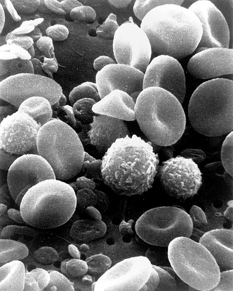
| 4 octobre 2006 à 03:00 |  | 1 800 × 2 239 (989 kio) | DO11.10 | {{Information |Description=This is a scanning electron microscope image from normal circulating human blood. One can see red blood cells, several white blood cells including lymphocytes, a monocyte, a neutrophil, and many small disc-shaped platelets. Red | |
| 4 octobre 2006 à 01:09 |  | 500 × 326 (36 kio) | DO11.10 | {{Information |Description= A three-dimensional ultrastructural image analysis of a T-lymphocyte (right), a platelet (center) and a red blood cell (left), using a Hitachi S-570 scanning electron microscope (SEM) equipped with a GW Backscatter Detector. |
Utilisation du fichier
Les 2 pages suivantes utilisent ce fichier :
Usage global du fichier
Les autres wikis suivants utilisent ce fichier :
- Utilisation sur ar.wikipedia.org
- Utilisation sur ar.wikiversity.org
- Utilisation sur ast.wikipedia.org
- Utilisation sur as.wikipedia.org
- Utilisation sur az.wikipedia.org
- Utilisation sur ba.wikipedia.org
- Utilisation sur be-tarask.wikipedia.org
- Utilisation sur be.wikipedia.org
- Utilisation sur bg.wikipedia.org
- Utilisation sur bn.wikipedia.org
- Utilisation sur bn.wikibooks.org
- Utilisation sur bs.wikipedia.org
- Utilisation sur ca.wikipedia.org
- Utilisation sur ce.wikipedia.org
- Utilisation sur ckb.wikipedia.org
- Utilisation sur cs.wikipedia.org
- Utilisation sur cv.wikipedia.org
- Utilisation sur cy.wikipedia.org
- Utilisation sur de.wikipedia.org
- Utilisation sur de.wikibooks.org
- Utilisation sur dv.wikipedia.org
- Utilisation sur el.wikipedia.org
- Utilisation sur el.wiktionary.org
- Utilisation sur en.wikipedia.org
Voir davantage sur l’utilisation globale de ce fichier.
